
In 2010 CFINs Core Experimental Facility was established. Our goal has been to establish a world class research facility where researchers can get access to all major modalities for investigating the human brain, help for study design and data analysis, as well as powerful IT infrastructure for data storage and analysis.
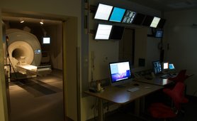
A Siemens TIM Trio MRI system was installed in January 2010. The scanner features both standard and advanced equipment, including: visual, auditory, electrical and thermal stimulation devices; monitoring devices for eyetracking, EMG, EOG, ECG, EEG, pulse oximetry, respiration and end-tidal CO2 and a number of devices to log subject responses. With lots of waveguides and an in-room monitor, the scanner room is extremely flexible and prepared for several experimental scenarios and optimized for both human and animal experiments.
In addition a Siemens Magnetom Skyra 3T was installed in October 2012, to replace our old General Electrics 3T MRI system. The Skyra scanner is equipped with the same accessories as mentioned above.
In December 2017 the Trio scanner was upgrade to a Prisma Fit scanner with more powerfull gradients, and better cooling.
In spring 2019 both scanners were moved to our new facility at Skejby Hospital, where they are placed next to testing and surgery rooms.
CFIN’s preclinical MRI lab is located at Aarhus’ new University Hospital in Skejby where it was moved to in Spring 2019. In its new location the preclinical MRI is located in rooms adjoining CFIN’s animal stable, micro surgery and optical labs ideally suited for all types of preclinical imaging projects and cross-modality investigations.
At the center of the preclinical MRI lab is a Bruker Biospec 9.4T horizontal small bore MR-system. The lab is fully equipped for a range of imaging studies from microimaging of biological samples to in vivo examinations. In vivo rodent brain imaging is the main interest for our activities. This is done using our cryo-array coil for rat brain offering superior sensitivity. Since the move to our current location the lab’s main focus has been to establish awake animal imaging. For this, we design and 3D print custom animal cradles so that our mouse studies also benefit from the boost in sensitivity offered by the rat cryo probe. It is the ambition of our lab to offer all types of MRI brain techniques for users.
Our lab is also well-equipped for microimaging of samples (predominantly fixed rodent brains but we have also imaged human bone, primate brain, porcine heart and brain, and the occasional whale teeth and eyes). Microimaging is performed using bore mounted room temperature coils ranging from 15-40 mm in diameter.
The preclinical MRI lab is headed by Dr Brian Hansen.

For information about possible projects and collaborations,
please contact Brian Hansen by email ...
In August 2011 we opened our Electa Neuromag TRIUX MEG scanner facility. The MEG scanner is placed below ground level at Aarhus University Hospital, close to the other research scanners, the researchers in Danish Neuroscience Center, and the clinical researchers at the hospital. The MEG scanner detects magnetic field changes in the femto-Tesla range, and therefore has to be isolated from outside sources such as moving vehicles and electrical fields. The scanner is being used for research within ultrafast brain processing, and for research based patient examinations prior to treatment of severe epilepsy.
During the summer of 2012, a Ultima In Vivo Two Photon Microscope from Prairie Technologies, including a Resonant Galvo Scanner, was installed in close proximity to other small animal research scanners (microPET and high field MR). The 2-photon microscope will be used for in vivo animal research in combination with oxygen probes and different stimulation paradigms. We expect to add equipment for FLIM and electrophysiological recordings in 2013.
